ZEISS Axiolab 5
Optimize Clinical and Biomedical Lab Efficiency with Smart Microscopy

Axiolab 5 meets the rigorous demands of your clinical and biomedical laboratories, experiencing the perfect blend of ergonomic design and speed. Profit from a completely new form of digital documentation with Axiolab 5 and the microscope camera Axiocam 208 color. Just focus your sample and press a single button for crisp images in true color. Smart Microscopy then takes over.

✓ Acquire digital images with a push of a button, that will look exactly like you see it through the eyepieces.

✓ Correct scaling information are added automatically.

✓ Work comfortably with adaptable ergonomics.

VIEW VARIANTS
Watch the ZEISS Axiolab 5 Product Video
ZEISS ZEISS Axiolab 5
Discover 10 highlights of Axiolab 5 in only 90 seconds.

Boost Your Efficiency

With Smart Microscopy

It can take a lot of time to acquire detail-rich, true-color images. You know the drill: place the sample, focus your region of interest, switch to the computer, adjust settings such as white balance, exposure time and gain, then acquire an image, insert a scale bar, switch back to the microscope … and so on.

That's what a typical documentation workflow looks like. Now, with the Axiolab 5 system, you can stay focused on your sample at all times, thanks to smart microscopy. Digital documentation is inherent in the system design. Just press the ergonomic Snap button on the microscope and you're done. The procedure integrates perfectly with your established microscopy workflow and boosts your efficiency tremendously.

How To Use
How-to Use a Barcode Reader with Smart Microscopy in Stand-Alone Mode

Faced with a high volume of samples or with sensitive data – or with both – uncomplicated, precise data management is key: you need to work quickly and with total data accuracy.

That’s where barcode labeling comes into its own, helping you work through even large sample quantities with great ease and reliability. Your smart microscope from ZEISS lets you assign microscope images with the correct scaling information to barcode-labeled samples. Just use your Axiolab 5 or Axioscope 5 microscope with Windows PC or iPad, connect a barcode reader to your Axiocam 208 color microscope camera and you’re good to go.

How-to Use Scan a Barcode with Labscope and Smart Microscopy

Faced with a high volume of samples or with sensitive data – or with both – uncomplicated, precise data management is key: you need to work quickly and with total data accuracy. That’s where barcode labeling comes into its own, helping you work through even large sample quantities with great ease and reliability.

Just use your Axiolab 5 or Axioscope 5 microscope with Axiocam 208 color camera and connect it with your iPad. By scanning the barcode with the built-in camera of the iPad, Labscope will assign that particular barcode-ID to the acquired image.

Ergonomics
In your clinical or biomedical lab, the ability to quickly evaluate patient samples and make informed decisions is of utmost importance. You constantly find yourself under time pressure, meticulously examining a vast number of tissue slides. As this work can be demanding, sitting in front of a microscope for long periods of time can take a toll on your comfort and well-being.

That's why achieving a comfortable and relaxed sitting position is crucial to your overall health. ZEISS ergotubes are engineered to ensure optimal ergonomic positioning, reducing strain and fatigue

Choose Between Three Configurations

With Axiolab 5 You Are Prepared for Today and the Future. Choose the Configuration, Which Suits Best to Your Lab Workflow:

  • Stand-alone for basic routine imaging

    Axiolab 5 in combination with Axiocam 202 mono or Axiocam 208 color operates independently of a computer system

  • ZEISS Labscope for advanced routine imaging

    Operating ZEISS Axiolab 5 with ZEISS Labscope imaging app is ideal for connected microscopy and standard multichannel fluorescence imaging.

  • ZEISS ZEN for research applications

    Use ZEN imaging software from ZEISS to perform additional processing and analysis of your images.

1 of 3
Related Applications

Microscopy Solutions for Cytology and Cytopathology

Detect Diseases on the Cellular Level

Cytopathology or cytology aims to detect diseases on the cellular level by the study of single cells and cell structures. This pathology discipline was founded in 1928 by the Greek medicine pioneer George Papanicolaou who also invented the “Pap smear”. These smear tests are now commonly used in cytopathology where clinical samples from a wide range of body sites are spread and deposited across a glass microscope slide, resulting in a thin film of cells for subsequent staining and examination by light microscopy.

Microscopy Solutions for Hematology

The Science or Study of Blood

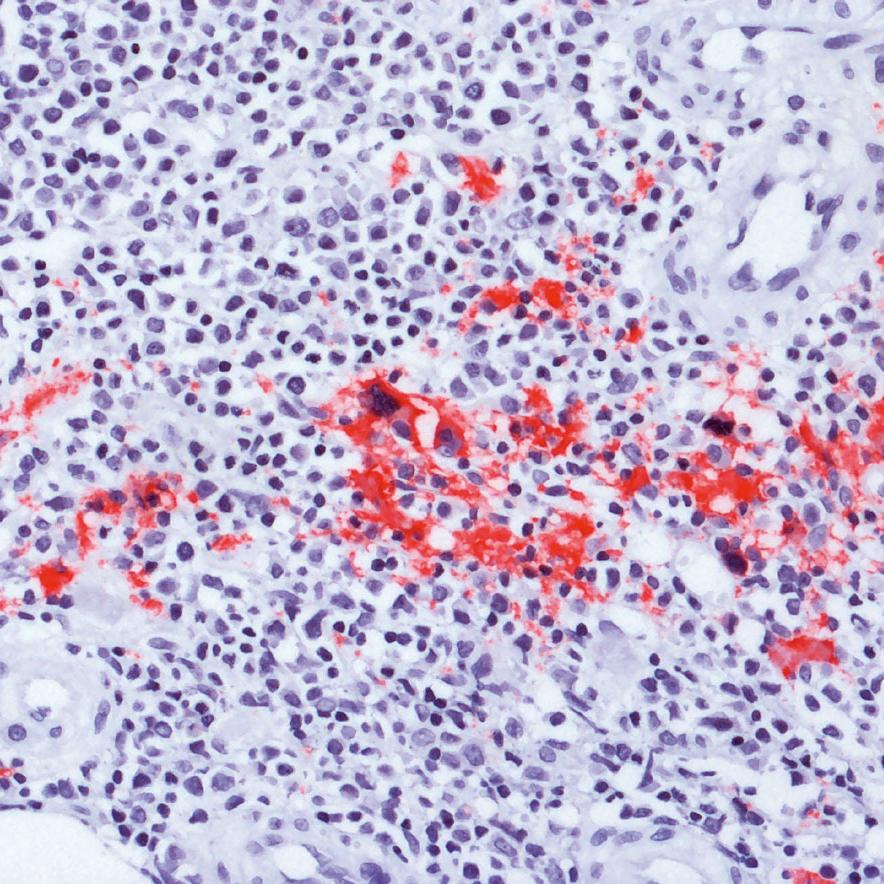
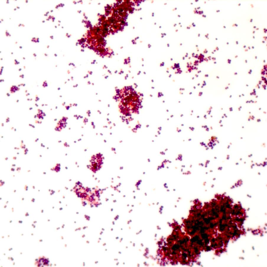
Hematology (alternate spelling “haematology”) is the science or study of blood, blood-forming organs and blood diseases. In clinical routine, hematologists diagnose and treat blood disorders and malignancies, including types of hemophilia, leukemia, lymphoma and sickle-cell anemia. Hematology is a branch of internal medicine that deals with the physiology, pathology, etiology, diagnosis, treatment, prognosis and prevention of blood-related disorders.

Blood consists of several components, including erythrocytes (red blood cells), leukocytes (white blood cells), thrombocytes (platelets) and plasma. All of the aforementioned cell types are produced in the bone marrow from multipotent precursor cells in a process called hematopoiesis.

Microscopy Solutions for Histology and Histopathology

Study the Manifestation of Disease by Microscopic Examination of Tissue Morphology

Pathology, histopathology or histology aims to study the manifestation of disease by microscopic examination of tissue morphology. In pathology, the sample to be examined under the microscope usually is the result of a surgery, biopsy or autopsy after fixation, clearing/embedding and sectioning of the tissue specimen. Alternatively, frozen section processing with a cryostat is done when rapid results are required (e.g. during surgery) or fixation would be detrimental to target structures such as lipids or certain antigens.

Microscopy Solutions for Microbiology

Prevention, Diagnosis and Treatment of Infectious Diseases

Microbiology is the branch of medical science concerned with the prevention, diagnosis and treatment of infectious diseases caused by pathogens such as bacteria, fungi, yeasts, parasites or viruses. A microbiologist studies the characteristics of pathogens, their modes of transmission, mechanisms of infection and growth to devise a treatment.

Microscopy Solutions for Semen Analysis

A Very Important Step Before Artificial Insemination

Semen analysis is a very important step before any artificial insemination is carried out. Semen is a fluid containing sperm cells, which is released during ejaculation. A normal sperm count is above 20 million sperm per milliliter of semen. The volume of semen is normally more than 2 milliliters.

Downloads

Product Info | White Papers

  • Product Info

    ZEISS Axiolab 5

    Your Smart Microscope for More Efficient Routine Lab Work

    DOWNLOAD
  • Product Info

    ZEISS Axiolab 5 - Flyer

    Your Smart Microscope for More Efficient Routine Lab Work

    DOWNLOAD
  • Product Info

    ZEISS Microscope for Cytopathology

    Detect Diseases on the Cellular Level

    DOWNLOAD
  • Product Info

    ZEISS Microscopes for Hematology

    Study Blood and Blood Diseases

    DOWNLOAD
  • Product Info

    ZEISS Microscopes for Histology

    Examine Tissue Morphologies

    DOWNLOAD
  • Product Info

    Your microscopes for your biomedical laboratory

    Your microscopes for increased efficiency in the lab.

    DOWNLOAD
  • White Papers

    Baumgartner – the Beer from the Innviertel Region

    Quality control with ZEISS Axiolab 5

    DOWNLOAD
  • White Papers

    Light Microscopic Analysis of Urine

    ZEISS Primostar 3 and ZEISS Axiolab 5

    DOWNLOAD
  • White Papers

    Microscopic analysis of pollen in honey

    Identification and documentation with ZEISS Axiolab 5

    DOWNLOAD
  • White Papers

    Microscopic Anatomy in the Study of Medicine

    Fundamentals of Histology

    DOWNLOAD
1 of 10