ZEISS AI Sample Finder
Reduce Your Time to Experiment From Minutes to Just Seconds​

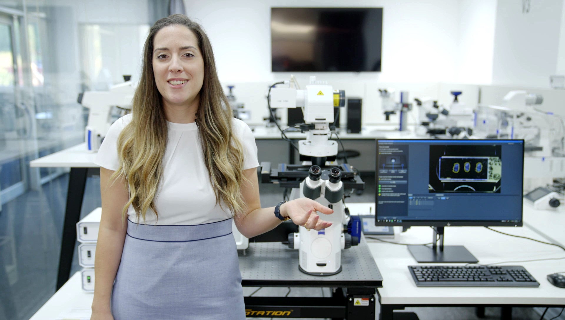
Microscopes are becoming increasingly automated. However, sample placement, focus adjustment and identification of the relevant sample areas still require manual adjustments. ZEISS AI Sample Finder automates this sequence entirely. After just seconds, you can access all sample areas directly and start your experiment right away.

✓ Automated sample placement

✓ Acquisition of an overview image

✓ Identification of the sample carrier

✓ Detection of relevant sample areas

✓ Focus adjustment for each region of interest


Get Support
Here Is How AI Sample Finder Works​

Automate the Start-up of Your Experiments

1. Sample placement

Place your sample on the loading position, the AI Sample Finder automatically moves it to the

2. Focus adjustment

The focus is adjusted automatically, and an overview image for fast and convenient navigation is taken within seconds. Composite darkfield illumination creates a high-contrast image even for very low-contrast samples.

3. Sample carrier identification

Intelligent routines automatically identify your sample carrier, regardless if you use a petri dish, a chamber slide, or a multi-well plate. Carrier properties are automatically transferred to the software, eliminating manual settings.

4. Sample detection

Deep Learning algorithms precisely detect even unusual sample regions. You can navigate and access all sample areas directly which allows you starting your experiment faster than ever.

See Your Entire Sample – Speed Up Your Microscopy Workflows

A good overview image is the foundation for a detailed analysis. AI Sample Finder enables you to see your entire sample with unmatched speed and ease of use. Forget about assignment issues after image acquisition. You will always know in which sample region your experiment was conducted and how the surrounding environment looked like. With ZEN Connect you can visualize to your data in a higher context combining different imaging modalities.

Overview image provided by AI Sample Finder, showing fluorescence, darkfield composite contrast, and a combination of fluorescence and coherence contrast (from left to right). Sample courtesy of M. Schmidt, Institute of Anatomy, Medical Faculty Carl Gustav Carus, Germany​